Photography
Better before photos, better afters
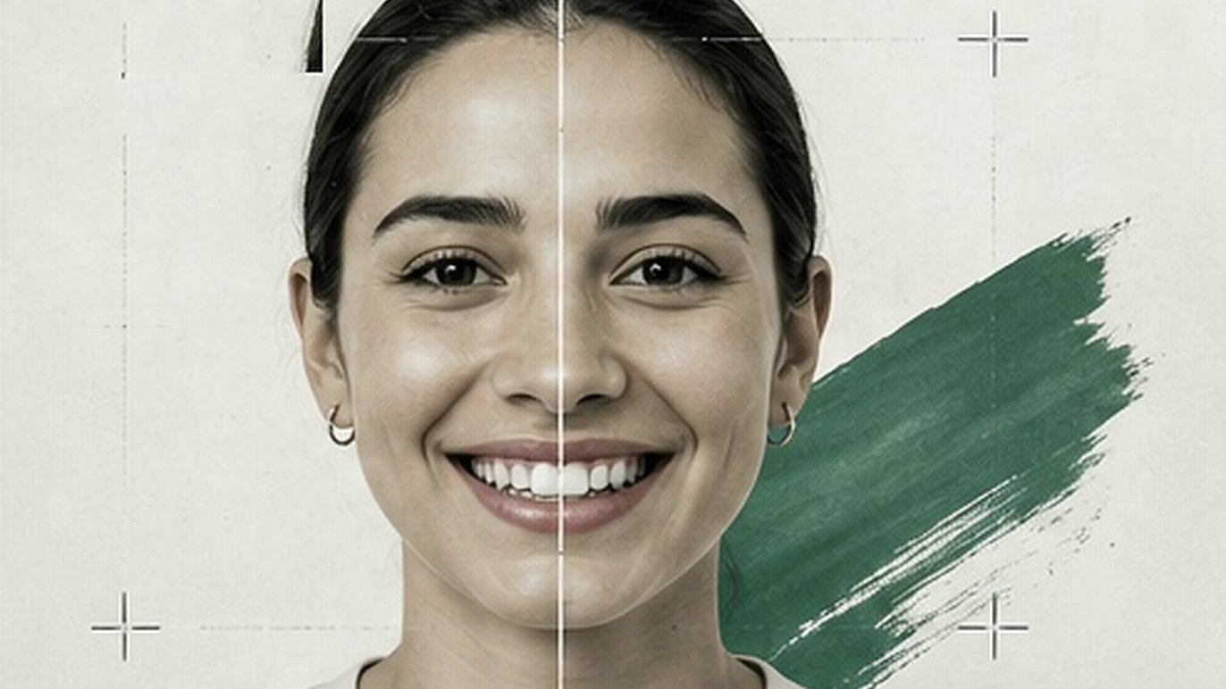
An orthodontist can spend two years producing an excellent result and still struggle to show it. Angle, lighting and expression decide whether the change is visible at all.
An orthodontist can spend two years producing an excellent result and still struggle to show it. Angle, lighting and expression decide whether the change is visible at all.
An orthodontist may spend 18 months or two years producing an excellent treatment result. Yet when the final photographs are placed beside the originals, the transformation can be surprisingly difficult to appreciate.
The problem may have nothing to do with the treatment.
The patient's head was slightly tilted in the first photograph. The camera was closer in the second. One was taken under bright daylight and the other under overhead office lighting. The patient gave a broad smile before treatment and a restrained one afterward. Or the original image simply wasn't sharp enough to stand beside the final photograph.
By the time anyone notices, the opportunity to fix it is long gone.
This is one of the peculiarities of before-and-after photography: the quality of the final comparison is largely determined on the day the first photograph is taken.
Photography is already an established part of orthodontic practice. Clinical photographs provide baseline records, assist with treatment planning and monitoring, support communication with patients and other clinicians, and have educational, research and medico-legal uses. The orthodontic literature consequently places considerable emphasis on producing images that are not only clear, but reproducible.
The growing use of treatment photography outside the clinical record adds another consideration. A well-documented case can help a patient understand what treatment achieved. With appropriate permission, photographs may also be used in case presentations, websites, educational materials and practice marketing. The ADA itself recommends high-resolution photographs of real patients in advertising where appropriate, while emphasizing the need for written permission.
The clinical photograph and the marketing photograph therefore increasingly occupy some of the same territory. But they are not necessarily the same thing.
# A Good Photograph and a Good Comparison Are Different Things
Modern cameras make it relatively easy to produce an attractive photograph. Producing a reliable before-and-after comparison is harder.
Imagine an adult orthodontic patient photographed before treatment with the camera slightly below eye level. At the end of treatment, the camera is positioned higher. Even if both photographs are individually excellent, differences in perspective and head position can change the apparent facial proportions and presentation of the smile.
Lighting creates similar problems. A darker initial photograph followed by a brighter, warmer final image can make teeth and skin appear different independent of treatment. Changes in camera distance, lens selection, cropping and patient expression can further exaggerate or conceal apparent differences.
This is why clinical photography literature emphasizes standardization of patient position, camera position, distance, lighting and background. Orthodontic photography guidance specifically describes reproducibility as important when photographs are used to document change over time.
For a practice interested in before-and-after photography, that leads to a useful principle: The objective is not to make the after photograph look better than the before photograph. It is to make the photography sufficiently similar that the treatment result speaks for itself.
That distinction matters clinically. It also matters for credibility.
# The Before Photograph Deserves More Attention
Practices naturally become interested in photography when a treatment result is ready to show. By then, half the work has already been done.
The before photograph establishes the composition that the practice will eventually need to reproduce. It determines the original camera angle, patient position, expression, framing, distance and lighting against which the final result will be judged.
A poor before photograph therefore creates a problem that cannot be completely solved by taking an exceptional after photograph.
The better approach is to photograph the patient at the beginning of treatment as though the practice already knows it will need to recreate that photograph at the end.
That means establishing a repeatable view rather than improvising each time. The patient's head position should be intentional. The amount of face or smile visible in the frame should be reasonably consistent. Camera distance and height should not change substantially. The background should be controlled, and the patient's expression should be natural enough that it can be reproduced later.
This does not require turning every photo session into a studio production. It requires removing unnecessary variation.
# Modern Smartphones and Tablets Can Produce Professional Patient Imagery
For many practices, the conversation about better photography quickly becomes a conversation about buying a better camera. That is increasingly unnecessary for many uses.
Modern smartphones can produce high-quality orthodontic photographs, and published research has specifically examined their use for orthodontic and dental clinical photography. A 2022 technique paper concluded that advances in smartphone cameras make high-quality orthodontic clinical photography possible when appropriate technique and supporting equipment are used.
More recent research gives a more detailed picture. A 2025 study in the European Journal of Orthodontics compared recent flagship smartphones with semi-professional DSLR cameras across a comprehensive set of intraoral and extraoral orthodontic photographs. DSLR systems performed significantly better for image quality and dimensional accuracy, although the study found no significant difference in ease of use or color-shade accuracy. The authors continued to recommend DSLR cameras with ring flash as the clinical gold standard.
Another 2025 study comparing an iPhone 14 Pro with DSLR and mirrorless systems similarly found the dedicated cameras superior overall. All DSLR and mirrorless photographs in that study were rated 'good,' compared with 41.7% of smartphone images; the remaining 58.3% of smartphone photographs were rated 'acceptable.' The authors concluded that smartphone images could be adequate for clinical records but cautioned against large-format applications.
Research published in 2026 adds an important qualification. In an in-vivo comparison involving 25 volunteers, three DSLR cameras and two recent flagship smartphones, researchers tested whether a standardized smartphone workflow could narrow the gap. With color calibration and appropriate optical zoom, smartphone photography achieved color accuracy and relative dimensional consistency comparable to DSLR cameras under the conditions studied.
Taken together, the evidence supports a more useful conclusion than either 'a phone is just as good as a professional camera' or 'professional dental photography requires a DSLR.' Modern smartphones and tablets can produce professional-quality patient imagery for many clinical communication and marketing purposes. Dedicated camera systems retain advantages where maximum standardization, macro photography, dimensional accuracy, controlled illumination or large-format output is required.
For most practices, the practical question is therefore not simply which camera has the best specifications. It is what the photographs need to accomplish.
# The Device Matters Less When the Process Is Repeatable
Smartphones and tablets have an obvious advantage in a busy practice: they are familiar.
A team member does not necessarily need to retrieve a camera from another room, attach a lens or remember a complicated set of controls before taking an extraoral photograph. Modern mobile devices are fast, accessible and capable of producing images with considerably more resolution than most websites or ordinary digital displays require.
But their automation can also introduce inconsistency. Modern phones may contain several lenses and switch between them depending on zoom level, distance and lighting. Automatic exposure can brighten one photograph more than another. Computational processing may alter contrast, color and sharpness. Moving the device closer to compensate for framing can change perspective.
A standardized mobile workflow therefore matters. Published guidance for smartphone orthodontic photography recommends deliberate lens selection, controlled patient positioning, appropriate camera distance, consistent lighting and a plain background. It also cautions against unnecessary digital zoom because of its effect on image quality.
The broader lesson applies regardless of device. A $3,000 camera used differently by every member of the team does not automatically create a reliable before-and-after record. A capable mobile camera used within a carefully defined process can produce much greater consistency.
Professional patient photography is not defined solely by owning a professional camera. It is also defined by using the camera professionally.
# Positioning Changes What the Viewer Sees
One of the most important variables in repeatable patient photography costs nothing. It is where the patient and camera are positioned.
For extraoral orthodontic photography, published technique guidance uses anatomical reference planes to establish reproducible patient positioning. For frontal views, for example, the interpupillary plane provides a reference; profile views can use the Frankfort plane.
A practice producing patient-facing before-and-after photographs does not necessarily need to turn those principles into an elaborate photographic procedure. But it should understand why they exist.
A slight upward or downward tilt of the head changes the presentation of the smile. Rotation changes facial symmetry. A camera held above the patient creates a different perspective from one held lower. Moving considerably closer with a wide-angle lens can change facial proportions.
If those variables differ between the before and after images, the viewer is no longer looking only at treatment change. They are looking at photographic change as well.
This is one reason the original photograph should be available when the final image is taken. Rather than relying on memory, the photographer can compare the new framing and position with the actual baseline.
# Lighting Should Reveal the Result, Not Create It
Lighting deserves similar attention.
The purpose of clinical before-and-after photography is not to make the final image as flattering as possible. It is to reveal the patient's appearance consistently.
Published orthodontic photography guidance emphasizes the importance of controlled lighting, while smartphone-specific guidance identifies lighting as one of the more challenging aspects of mobile dental photography and discusses the use of external continuous lighting where needed.
For a practice, consistency can be more important than complexity. Using the same area of the practice, a similar background and similar lighting conditions removes variables from the comparison. Strong daylight in one photograph and dim artificial lighting in another can change exposure, color and shadow. The camera's automatic processing may then amplify those differences.
The final photograph may be beautiful. But a beautiful photograph is not necessarily an honest comparison.
# A Natural Expression Is Part of Standardization Too
Cameras are not the only variable. Patients are.
Ask someone to smile twice and the expressions will rarely be identical. Ask for the 'biggest possible smile' and the variation can become even greater.
For patient-facing before-and-after photography, a relaxed, natural smile is generally easier to reproduce and often more representative of how the patient actually looks.
The instruction matters as well. If one team member says 'smile naturally' and another encourages a patient to smile as broadly as possible, the practice has introduced another variable into the comparison.
A simple, consistent instruction can make the photographs more repeatable. Taking several frames also makes sense. Storage is inexpensive. The opportunity to photograph the pretreatment condition again after treatment has begun does not exist.
# The After Photograph Should Recreate, Not Reinvent
When treatment is complete, there is a natural temptation to treat the final photograph as a new photo session. For a before-and-after comparison, it is better thought of as a recreation.
Open the original photograph. Look at how the patient was positioned. Compare the amount of face visible in the frame. Look at head rotation, camera height, background and expression. Then reproduce those conditions as closely as reasonably possible.
The goal is not pixel-perfect duplication. Patients grow, hairstyles change, equipment changes, practices move rooms and treatment itself may affect facial appearance.
The goal is to eliminate the differences the practice can control.
This is especially important when photography is distributed among several team members. Without a standard process, the before image may be taken by one person and the after image by someone else 18 months later, each with a different idea of what a good patient photograph looks like.
Standardization gives both photographers the same target.
# Clinical Records and Marketing Assets Are Not the Same Thing
A photograph can be clinically useful without being suitable for a practice website. The reverse can also be true.
A highly polished portrait may look excellent in marketing but omit views or details necessary for clinical documentation. A standardized intraoral image may be important for diagnosis and records while being of little value as patient-facing marketing.
Practices should therefore decide what each photographic workflow is intended to produce.
There will sometimes be overlap. A well-composed extraoral before-and-after image can be useful for clinical documentation, patient communication and, with appropriate authorization, marketing.
But using the same image for a different purpose changes the privacy and consent considerations surrounding it. This is particularly important in the United States.
# A Patient Photograph Can Be Protected Health Information
The ADA cautions dental practices that protected health information under HIPAA can extend beyond conventional patient records. A photograph or video can constitute protected health information when it identifies the patient or contains sufficient information to identify them.
That means a practice should not assume that removing a patient's name automatically makes a photograph appropriate for marketing.
The ADA advises practices to obtain written permission before using real-patient photographs in advertising. It also states that HIPAA-covered dental practices need valid written authorization before using full-face photographs and comparable identifiable images for marketing.
HHS guidance provides the broader regulatory context: under the HIPAA Privacy Rule, communications falling within its definition of marketing generally require the individual's authorization, subject to specified exceptions.
Practices should determine the exact requirements applicable to their circumstances, including federal and state law, rather than treating a generic release as legal advice.
The practical lesson is simpler: consent for treatment photography and authorization to publish a patient's image should not be treated as the same decision.
A practice may have a legitimate clinical reason to photograph a patient without having permission to place that photograph on social media or its website.
# Before-and-After Photography Should Not Make the Result Better Than It Was
Digital photography also creates a less obvious ethical question: how much should an image be edited?
Some post-processing is routine. Cropping an image, correcting orientation or making reasonable adjustments to exposure can help produce consistent presentation.
But the purpose of a treatment comparison changes if editing begins to alter the treatment result itself.
Whitening teeth digitally, changing gingival contours, removing clinical imperfections, altering facial proportions or modifying other relevant anatomy can turn documentation into something else. Even aggressive differences in exposure, contrast and color can make an after image appear more dramatic than the original result.
The basic advertising principle is not complicated: the image should not mislead the viewer about what treatment achieved. The ADA notes that dental advertising remains subject to obligations concerning fairness and accuracy regardless of whether it appears in traditional or newer media.
AI makes that distinction more important, not less. Image-editing systems can now make sophisticated changes in seconds that previously required substantial retouching expertise. The fact that a modification is technically easy does not make it appropriate for clinical or before-and-after documentation.
A credible before-and-after comparison should help the viewer see the result more clearly. It should not manufacture a better one.
# Standardization Is a Practice System, Not a Photography Hobby
There is a tendency for dental photography to become the responsibility of the person in the practice who happens to be interested in cameras.
That can produce excellent photographs. It can also create a fragile system.
If patient photography is genuinely useful to the practice, the process should work when that person is on vacation, when a new assistant joins the team or when the practice changes devices.
Research suggests that this lack of standardization remains a real issue. A 2025 survey of 304 orthodontists in Spain found substantial variation in photographic equipment and practices. DSLR cameras with macro lenses and ring flashes were the most commonly used setup, but the study also found considerable interest in additional photography training and concluded that standardized protocols could improve clinical documentation and diagnostic consistency.
A practical protocol does not need to be complicated. It needs to establish what views are required, how the patient is positioned, where the camera is placed, what lens or mobile camera mode is used, what should appear in the frame, how lighting and background are controlled, when photographs are taken, where they are stored and how the final image is matched with the original.
Most importantly, someone should look at the photograph before the patient leaves. A blurry pretreatment image discovered six months later cannot be retaken.
# The Best Before-and-After Photograph Is the One You Can Trust
Patient photography occupies an interesting position in modern orthodontics.
It remains part of the clinical record and professional documentation. At the same time, patients increasingly encounter photographs when researching practices, understanding treatment and evaluating what orthodontic care can achieve.
Modern smartphones and tablets make high-quality photography far more accessible than it once was. Dedicated DSLR and mirrorless systems continue to offer advantages for demanding clinical applications, but professional patient imagery no longer begins and ends with ownership of a professional camera. The evidence increasingly shows that technique, distance, lens choice, lighting and standardization materially influence what a mobile camera can produce.
For before-and-after photography, that leads back to the simplest point.
The practice should not try to make the after photograph more impressive than the before. It should try to make everything except the treatment as similar as possible.
Same type of view. Similar camera position. Similar distance. Similar head position. Similar lighting. Similar expression. A clear original image preserved as the reference for the final photograph.
When those conditions are controlled, the photography becomes quieter. And the treatment result becomes easier to see.
The most important photograph in a before-and-after pair may therefore not be the one everyone wants to show. It is the one taken before anything has changed.
# Data and Methodology
This article draws on peer-reviewed research in orthodontic and dental photography together with guidance from the American Dental Association and U.S. Department of Health and Human Services. Practical workflow considerations were also informed by SmileStudio's supplied before-photo guidance, which emphasizes obtaining a usable, reproducible baseline image rather than treating photography as an improvised step.
Smartphone photography evidence: Published studies do not establish that smartphones and DSLR cameras are universally interchangeable. A 2025 European Journal of Orthodontics study found DSLR systems superior for image quality and dimensional accuracy. A separate 2025 comparison found smartphone photographs ranging from good to acceptable but inferior overall to DSLR and mirrorless systems. A 2026 in-vivo study found that under a standardized workflow using color calibration and appropriate optical zoom, recent flagship smartphones could achieve color and relative dimensional consistency comparable to DSLR systems under the conditions tested.
Clinical versus marketing use: Evidence supporting smartphone photography for clinical documentation should not automatically be interpreted as evidence of suitability for every commercial output. Requirements differ according to intended use, including image size, diagnostic purpose and presentation medium.
Privacy and authorization: ADA and HHS materials are used to describe general U.S. privacy and marketing considerations. They are not a substitute for legal advice, and practices should evaluate HIPAA, applicable state laws and their individual circumstances before capturing, disclosing or publishing patient imagery.
Important limitation: Camera technology changes rapidly. Research comparing a specific smartphone generation with DSLR or mirrorless systems should not be interpreted as a permanent ranking of all smartphones and dedicated cameras.
# Primary and Supporting Sources
Peer-reviewed - Technique for Orthodontic Clinical Photographs Using a Smartphonehttps://pmc.ncbi.nlm.nih.gov/articles/PMC8800594/
European Journal of Orthodontics - Smartphones or digital SLRs for clinical dental photography: is there a difference?https://pubmed.ncbi.nlm.nih.gov/40501276/
Journal of Orthodontics - Does the camera type affect the quality of orthodontic photographs?https://pubmed.ncbi.nlm.nih.gov/40819226/
Clinical Oral Investigations - Comparison of accuracy and reliability between smartphone and DSLR photographyhttps://pubmed.ncbi.nlm.nih.gov/42461427/
Peer-reviewed - Techniques for Capturing Clinical Photographs in Orthodonticshttps://pmc.ncbi.nlm.nih.gov/articles/PMC11645108/
American Dental Association - Traditional Advertising and Patient Photographyhttps://www.ada.org/resources/practice/practice-management/38_otheradvertising_traditional
American Dental Association - Marketing and Advertisinghttps://www.ada.org/resources/practice/legal-and-regulatory/marketing-and-advertising
American Dental Association - Sample Photography/Image Releasehttps://www.ada.org/-/media/project/ada-organization/ada/ada-org/files/publications/guidelines-for-practice-success/gps-marketing/sample-photography_image-release.pdf
U.S. Department of Health and Human Services - HIPAA and Marketinghttps://www.hhs.gov/hipaa/for-professionals/privacy/guidance/marketing/index.html
Last updated
August 2026
See it on your own casesTwenty minutes, on a call, with your own cases in front of you.
Schedule a free demo